


In Spain, epidemiological studies of the prevalence of diffuse interstitial lung disease (ILD) in rheumatoid arthritis (RA) are limited. Our objective was to estimate the prevalence of symptomatic ILD in RA and its characteristics in our area.
Materials and methodsIn our hospital’s interdisciplinary rheumatology and pulmonology clinic, a prospective longitudinal observational study was designed in which we included RA with respiratory symptoms and ILD confirmed by high resolution computed tomography.
ResultsOf the 2729 people with RA in our area, 47 had symptomatic ILD, estimating a prevalence of symptomatic ILD in RA of 1.72% (95% CI 1.26–2.29) with an age at diagnosis of RA of 57.3 ± 13.3 years. It was more frequent in men, 60.6% had a history of smoking, and 84.3% and 84.7% had rheumatoid factor (RF) and anti-cyclic citrullinated peptide (Anti-CCP) antibodies, respectively. The most frequent pattern was usual interstitial pneumonitis (UIP), appearing in 28 (31.1%). Nonspecific interstitial pneumonia (NSIP) was more frequent in women, while the combined pulmonary fibrosis-emphysema (CPFE) syndrome presented exclusively in men.
ConclusionsWe have analysed the prevalence of symptomatic RA-ILD in our area, which is lower than expected, probably in relation to the definitions used. We have also described that the UIP pattern is the most frequent in RA in our environment, followed by the NSIP. Lastly, we have analysed the prevalence of CPFE in RA, which reaches 13%, for the first time.
En España, los estudios epidemiológicos de prevalencia de enfermedad pulmonar intersticial difusa (EPID) en artritis reumatoide (AR) son escasos y limitados. Nuestro objetivo fue estimar la prevalencia de EPID sintomática en AR y sus características en nuestra área.
Materiales y métodosSe diseñó un estudio observacional longitudinal prospectivo en la consulta interdisciplinar de Reumatología y Neumología, en el que incluimos AR con síntomas respiratorios y EPID confirmada por tomografía computarizada de alta resolución.
ResultadosDe las 2.729 personas con AR de nuestra área, 47 presentaban EPID sintomática, estimándose una prevalencia de EPID sintomática en AR del 1,72% (intervalo de confianza del 95%: 1,26−2,29) con una edad al diagnóstico de AR de 57,3 ± 13,3 años. Fue más frecuente en hombres, el 60,6% tenía antecedente de tabaquismo y el 84,3 y el 84,7% factor reumatoide y anticuerpos antipéptidos cíclicos citrulinados, respectivamente. El patrón más frecuente fue neumonía intersticial usual (NIU) en 28 (31,1%), la neumonía intersticial no específica (NINE) fue más frecuente en mujeres y el síndrome combinado enfisema-fibrosis (SCEF) exclusivamente en hombres.
ConclusionesEn este estudio hemos analizado la prevalencia de AR-EPID sintomática en nuestra área, la cual está por debajo de lo esperado, probablemente en relación con las definiciones utilizadas. Así mismo, hemos descrito que el patrón NIU es el más frecuente en la AR, seguido del NINE y analizado por primera vez la prevalencia de SCEF en la AR, que alcanza el 13%.
Rheumatoid arthritis (RA) is a systemic inflammatory disease characterised by chronic inflammation of the joints and their resulting destruction, deformity, and incapacity1, with extra-articular manifestations that contribute to morbidity and mortality2. The lungs are a commonly affected extra-articular location, with diffuse interstitial lung disease (ILD) representing the most frequent manifestation3. Up to 58% of patients with RA may experience lung involvement, which is associated with mortality in 10%–20% of cases4.
The prevalence of clinical RA with ILD (RA-ILD), its risk factors, and its presentation are of great interest. In Spain, there are very few epidemiological studies on the prevalence of RA-ILD, and these are limited5, and descriptive studies on lung involvement include few patients with RA6,7.
In 2007, a pioneering interdisciplinary consultation unit was set up at the La Paz University Hospital (HULP) in Madrid by the Rheumatology and Pulmonary services to jointly assess lung involvement in immune-mediated systemic diseases. The consultation unit was integrated with the centre’s ILD unit, accredited by the Spanish Society for Pulmonology and Thoracic Surgery (SEPAR) as a highly complex interdisciplinary unit. This made it possible to collect data in a structured manner, enabling all the dimensions of the disease to be studied within our area.
The objective of this study was to estimate the prevalence of symptomatic ILD, its radiological patterns in RA, and the characteristics of patients who develop respiratory symptoms within the health district of a tertiary referral hospital.
Patients and methodsA prospective, longitudinal, observational study was designed between 2007 and 2018 in the HULP Rheumatology and Pulmonary interdisciplinary consultation unit. The protocol was approved by the HULP Research Ethics Committee.
PopulationWe included patients from the health district area and those seen in the Rheumatology service who met the RA criteria from 1987 or from 20108,9 and those who had been referred to the interdisciplinary Rheumatology and Pulmonary consultation unit with suspected ILD due to symptoms (cough, dyspnoea), signs (pathological lung sounds), or alterations in imaging tests.
Case definition and data collectionSymptomatic ILD was defined in the presence of cough, dyspnoea, or crackles in pulmonary auscultation in patients with high-resolution computed tomography (HRCT) diagnostic of ILD. Patients with overlap syndrome or other possible causes of ILD were excluded.
Sociodemographic and clinical variables were collected, as well as data regarding smoking, received RA treatments, respiratory symptoms (cough and dyspnoea), presence of crackles, rheumatoid factor titre, and cyclic citrullinated peptide antibodies (CCPA), pulmonary function tests (PFTs), and HRCT radiological patterns.
MasterLab-body 6.0 equipment (Viasys, Wuerzburg, Germany) was used for the PFTs according to the ATS/ERS recommendations for the calibration and measurement procedures10. The procedure for slow and forced spirometry was that described by SEPAR11. The values proposed by the Global Lung Initiative were used as reference values12. The diffusing capacity for carbon monoxide (DLCO) was measured using the same equipment with the single breath technique and was corrected according to haemoglobin, with the reference values proposed by Cotes13. Pulmonary function was considered abnormal if the FVC or DLCO were under 80%14.
ILD diagnosis was made using HRCT; the protocol included a topogram to determine the volume limits that should be acquired in maximal inspiration, in the craniocaudal direction. Slices during expiration at the arch and at the entry to the pulmonary veins were also obtained to assess air entrapment. A 16-detector SOMATOM EMOTION 16 CT scanner was used (Siemens Medical Solutions, Erlangen, Germany).
The irradiation parameters were adjusted conventionally according to the morphological characteristics of each patient: 120 kV, with tube current of 160 mA. The slice thickness used was 0.75 mm, with a reconstruction increment of 0.5 mm, a pitch ratio of 0.8 mm and a collimation thickness of 0.6 mm. Two standard reconstruction algorithms were used (filter B 41 to assess soft tissues and filter B 90 in high resolution for better evaluation of the lung parenchyma.
The images were interpreted by two radiologists from the chest service of our hospital, experts in interstitial disease and part of the multidisciplinary ILD team.
Statistical analysisThe sociodemographic and clinical data, the PFTs, and radiological patterns were analysed. The sample description was performed using measures of central tendency and dispersion for quantitative variables, as well as frequency tables and percentage distributions for the qualitative variables.
After describing the analysis sample, the population prevalence of symptomatic ILD in RA was estimated in the cases belonging to the reference area for HULP.
For the ILD prevalence study, the denominator was considered all the RA cases provided by the health centres in the area who were alive in October 2018, while those who presented with symptomatic ILD were the numerator. A Poisson distribution was assumed to calculate the prevalence and their 95% confidence interval (CI). The results were expressed globally and by age and sex strata.
For the patients with ILD, the frequency was established for the different radiological patterns classified as: usual interstitial pneumonia (UIP); probable UIP; combined pulmonary fibrosis-emphysema syndrome (CPFE); nonspecific interstitial pneumonia (NSIP); organizing pneumonia (OP); lymphoid interstitial pneumonia (LIP), and others. To do this, we used the idiopathic interstitial pneumonias classification from the European Respiratory Society (ERS) and the American Respiratory Society from 2013, and the recommendations for idiopathic pulmonary fibrosis drafted jointly by the American Thoracic Society, the European Respiratory Society, the Japanese Respiratory Society, and the Latin American Thoracic Society15,16.
The following pattern groups were created and compared: (UIP + probable UIP) vs. (NSIP).
We compared the clinical and sociodemographic characteristics as well as the baseline PFTs between both groups using statistical hypothesis testing for continuous variables (student’s t-test or Mann–Whitney U test, according to the distribution) and categorical variables (chi-squared).
ResultsA total of 2729 cases of RA were recorded in the area, which represents a prevalence of RA of 0.62%, which is to be expected. The symptomatic RA-ILD registry included 92 patients and 2 were eliminated (one case of juvenile idiopathic arthritis and one of pneumoconiosis). Of the 90 patients included, 69 belonged to the HULP district and 47 were alive as of the study date.
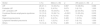
The population prevalence of symptomatic RA-ILD in the HULP health district was estimated to be 1.72% (95% CI: 1.26–2.29). By sexes, it was higher in males (3.11% vs. 1.23%), by age groups, the highest value was obtained in those younger than 55 years compared to those older than this age (2.20 95% CI: 1.28–3.52 vs. 1.53, 95% CI: 1.03–2.19). Although the CIs do not overlap for sex, which indicates the difference is statistically significant, this is not true for age and the observed difference could be due to chance (Table 1).
Prevalence of diffuse interstitial lung disease in rheumatoid arthritis, area pertaining to Hospital Universitario La Paz (Madrid).
| ILD, n | Prevalence (95% CI) | |
|---|---|---|
| Global (denominator: 2729 RA area) | 47 | 1.72 (1.26−2.29) |
| Per sex | ||
| Male (n = 706) | 22 | 3.11 (1.95–4.72) |
| Female (n = 2023) | 25 | 1.23 (0.80–1.82) |
| Per age group | ||
| <55 years (n = 774) | 17 | 2.20 (1.28–3.52) |
| ≥55 years (n = 1955) | 30 | 1.53 (1.03–2.19) |
CI: confidence interval; ILD: diffuse interstitial lung disease; RA: rheumatoid arthritis.
Table 2 shows the description of the 90 patients with RA and ILD in our series. Mean age at RA diagnosis was 57.3 ± 13.3 years and 49 (48.9%) were female. The majority, RF and positive CCPA, 16% smokers and 45% ex-smokers. Articular involvement was the initial symptomatology in 80 cases (90%), with an average time elapsed between onset of articular symptoms and respiratory symptoms of 9.9 years. At the baseline visits, 23% presented with cough; 58% with dyspnoea; 15.2% with cough and dyspnoea; 66% with crackles.
Description of the sample of patients with rheumatoid arthritis and diffuse interstitial lung disease, area pertaining to Hospital Universitario La Paz (Madrid).
| Characteristic | No. of patients studied | Results |
|---|---|---|
| Belonging to the area, n (%) | 90 | 69 (76.7) |
| Female sex, n (%) | 90 | 44 (48.9) |
| Age at diagnosis, m ± SD (years) | 90 | 57.3 ± 13.3 |
| Positive RF, n (%) | 89 | 75 (84.3) |
| RF titre, m ± SD | 89 | 688.8 ± 1184 |
| Positive CCPA, n (%) | 85 | 72 (84.7) |
| CCPA titre, m ± SD | 85 | 1011.1 ± 1045 |
| Smoking, n (%) | 89 | |
| Non-smoker | 35 (39.3) | |
| Ex-smoker | 40 (44.9) | |
| Smoker | 14 (15.7) | |
| Initial symptomatology, n (%) | 90 | |
| Joint | 80 (89.9) | |
| Pulmonary | 10 (11.1) | |
| Time of articular-pulmonary symptoms (years), m ± SD | 80 | 11.3 ± 9.2 |
| Time of pulmonary-articular symptoms (years), m ± SD | 10 | 1.1 ± 1.4 |
| Pulmonary signs and symptoms, n (%) | 86 | |
| No cough or dyspnoea | 29 (33.7) | |
| Cough or dyspnoea | 44 (51.16) | |
| Cough and dyspnoea | 13 (15.2) | |
| Crackles | 86 | 57 (66.3) |
| DLCO % (first measurement), m ± SD | 69 | 0.73 ± 0.21 |
| FVC % (first measurement), m ± SD | 69 | 0.93 ± 0.19 |
| Start of disease-start of treatment (years), m ± SD | 86 | 1.2 ± 3.3 |
| Start of disease-start of treatment (months), m ± SD | 86 | 15.0 ± 40.5 |
| Death, n (%) | 84 | 26 (30.9) |
CCPA: cyclic citrullinated peptide antibody; DLCO: diffusing capacity for carbon monoxide; FVC: forced vital capacity; RF: rheumatoid factor; SD: standard deviation.
The most common radiological pattern was UIP (31%), followed by NSIP (22%), CPFE (13%), the nonspecific pattern (10%), and OP and LIP (6.7%) (Table 3). Regarding the distribution by sex, NSIP was more frequent in females (34% vs. 13%; p = 0.032) and CPFE only appeared in males (26%; p < 0.0001). Regarding age, a higher frequency of NSIP was observed in those <55 years (32% vs. 14%; p = 0.048) and CPFE in the oldest age group (20% vs. 5%; p = 0.03). UIP was also more common in those older than 55 years (39% vs. 22%; p < 0.08) but did not reach statistical significance (SS) (Table 3).
Description and distribution of radiological patterns of diffuse interstitial lung disease in global rheumatoid arthritis (n = 90), by sex and age group.
| Global | n (%) | Male (n = 46) | p | ≥55 years (n = 49) | p |
|---|---|---|---|---|---|
| UIP | 28 (31.1) | 14 (30.4%) | 0.887 | 19 (38.8%) | 0.086 |
| Probable UIP | 5 (5.6) | 2 (4.3%) | 0.673 | 3 (6.1%) | 1.000 |
| UIP + probable UIP | 33 (36.3) | 16 (34.8%) | 0.705 | 22 (44.9%) | 0.076 |
| Combined fibrosis-emphysema syndrome | 12 (13.3) | 12 (26.1%) | <0.0001 | 10 (20.4%) | 0.033 |
| NSIP | 20 (22.2) | 6 (13.3%) | 0.032 | 7 (14.3%) | 0.048 |
| Organizing pneumonia | 6 (6.7) | 2 (4.3%) | 0.429 | 1 (2.0%) | 0.054 |
| Lymphoid interstitial pneumonia | 6 (6.7) | 1 (2.2%) | 0.107 | 3 (6.1%) | 1.000 |
| Others | 13 (14.4) | 9 (10%) | 0.489 | 1 (2.3%) | 1.000 |
NSIP: nonspecific interstitial pneumonia; UIP: usual interstitial pneumonia.
In the comparison by radiological groups, UIP + probable UIP versus NSIP (Table 4), the mean age at diagnosis in the group with UIP + probable UIP was 64.2 versus 52.3 (p = 0.039) and NSIP was more common in females (70% vs. 51.5%; p = 0.186) but did not reach SS (p = 0.18). When comparing the NSIP pattern with UIP + probable UIP, it was observed that the RF titre was greater in patients with NSIP (p = 0.020). Regarding DLCO and FVC, we did not find any significant differences, likely due to the sample size (Table 4).
Comparison of radiological patterns.
| UIP, probable UIP (n = 33) | NSIP (n = 20) | p | |
|---|---|---|---|
| Age at diagnosis (years) | 64.2 (54.0−69.0) | 52.3 (44.8−64.1) | 0.039 |
| Female sex, n (%) | 17 (51.5) | 14 (70.0) | 0.186 |
| Positive RF, n (%) | 28 (84.8) | 20 (100) | 0.144 |
| RF titre | 211 (45−766) | 620 (236−1135) | 0.020 |
| Positive CCPA, n (%) | 24 (82.8) | 20 (100) | 0.070 |
| CCPA titre | 624 (56−1256) | 1350 (312−1812) | 0.179 |
| Smoking, n (%) | 0.652 | ||
| Ex-smoker | 14 (43.7) | 6 (30.0) | |
| Smoker | 3 (9.4) | 2 (10.0) | |
| DLCO (first) | 0.73 (0.59–0.91) | 0.68 (0.54–0.82) | 0.526 |
| FVC (first) | 0.85 (0.77–0.95) | 0.94 (0.78−1.09) | 0.282 |
| Rheumatoid nodules, n (%) | 4 (13.3) | 1 (5.3) | 0.636 |
| Secondary Sjogren’s syndrome, n (%) | 3 (10.0) | 2 (10.5) | 1.000 |
| Carpal tunnel, n (%) | 1 (3.3) | 1 (5.3) | 1.000 |
| Death, n (%) | 13 (41.9) | 6 (31.6) | 0.464 |
Continuous variables expressed as medians (P25–75).
CPA: citrullinated peptide antibodies; DLCO: CO diffusing capacity; FVC: forced vital capacity; NSIP: nonspecific interstitial pneumonia; RF: rheumatoid factor; UIP: usual interstitial pneumonia.
Statistically significant data are written in bold (p < 0.05).
In our study, a prevalence of ILD in RA lower than that described in other series was estimated17, thereby confirming that this disease is more frequent in smokers and patients with RF and positive CCPA and that UIP is the most frequent pattern.
Although the study is from a single centre, it involves a large representative area in which almost all RA patients are referred to Rheumatology (with an RA prevalence of 0.62 [95% CI: 0.60–0.64] according to own data, which matches that estimated for the general population). In addition, patients seen in the interdisciplinary consultation unit were referred by both the hospital’s Rheumatology service as well as specialised centres, meaning patients with RA across the entire spectrum of disease severity were included. Lastly, their clinical characteristics are very similar to those of other series, which supports their representation.
The prevalence of ILD in patients with RA varies from 1% to 67.3%17. These differences are not surprising due to the large number of definitions, diagnostic tests used18, criteria19, and even due to the statistical analysis used, which is not always reflected in articles19. The prevalence ranges from 1% to 6% if chest X-ray is used and from 5% to 67.3% if HRCT is performed20.
It must be considered that, while the proportion of patients with RA-ILD via HRCT is noteworthy, it is only considered clinically significant in 5%–10%21. This must be taken into account given that there is a significant gap between the large number of asymptomatic individuals with functional or radiological pulmonary changes and the number of patients who ultimately develop clinical ILD22. Kelly et al.23 conducted a study in symptomatic patients diagnosed via HRCT and obtained a result of 2%–3%. Likewise, in a study including patients with pleurisy or ILD, Bartels et al. published results of 2% in 1985 and 4% in 200618.
In Spain, the only available data on the prevalence of ILD in RA are those from the EMECAR cohort5, with a prevalence of 3.7% (95% CI: 2.4–5). In this study, the classification criteria consisted of the existence of a restrictive pattern or HRCT findings indicative of ILD but without any reference to the symptoms. Despite the fact that descriptive studies of RA-ILD in our country have recently been published6,7, we have not identified other studies that estimate its prevalence.
The prevalence of symptomatic RA-ILD in our area is 1.72% (95% CI: 1.26–2.29), comparable to the results of EMECAR5 and to the work of Kelly et al.23 Our study only includes patients with respiratory symptoms for whom we have confirmed ILD via HRCT. This means that our study could have underestimated the prevalence of RA-ILD. In addition, the fact that we did not select for only patients with severe RA could also explain this low prevalence.
In RA, respiratory involvement is the first manifestation in 10%–20% of patients3, data that coincide with our results, wherein 10 patients (10%) initially presented with respiratory symptoms. Onset of respiratory involvement in RA typically occurs around 50–60 years24 and age is considered a risk factor for developing ILD. On the other hand, age at diagnosis of RA is also related to ILD22. In our series, the mean age at diagnosis of RA was 57 years. It is noteworthy that the prevalence of ILD is higher in those younger than 55 years rather than those older, though these differences are not statistically significant.
In addition to age, smoking, RF and positive CCPA, particularly at high titres, family history of RA, and being male have been described as risk factors for developing ILD with RA23,25–27. In our series, 61% of the patients had a history of smoking and over 80% had RF and positive CCPA at high titres, which fits with the expectations.
In immune-mediated systemic diseases, the NSIP pattern is the most common with the exception of RA, for which UIP is the most frequent19. In our study, UIP is the most frequent (31.1%), followed by NSIP (22.2%), with a pattern similar to other publications, with numbers in the range of 40%–60% for UIP and 11%–30% for NSIP4. There are increasingly more data confirming that patients with RA and UIP pattern have a different phenotype than patients with non-UIP patterns. UIP is more common in males, older patients, and smokers3, while the NSIP pattern is almost 3 times more common in females28. As in other works, patients with UIP or probable UIP are older at diagnosis (p = 0.039), are more commonly men (48.5% vs. 30%), and more frequently have a history of smoking than the NSIP group (53.1% vs. 40%).
The majority of studies on ILD in auto-immune diseases have not taken in account the coexistence with emphysema, meaning the frequency of CPFE in these diseases is not known29. In our study, 13.3% of the patients showed a CPFE pattern. We were also able to confirm that these patients have a special phenotype; in our study, all were male, smokers, and over the age of 55.
The study presents some limitations. We used the age stratification provided by the primary care registries, which could represent a limitation given that the limit of 55 years is different from other series and does not allow for comparisons. Although the study was conducted at one centre, we think that the sample is sufficiently representative for the data to be extrapolated to other areas. The fundamental strength of the study is that the patients were recruited from an interdisciplinary unit which featured a diagnosis and follow-up protocol, allowing us to collect highly uniform data.
ConclusionsWith this single-centre study, we have analysed the prevalence of symptomatic RA-ILD in our area, which is under 2%, this being far lower than that described in other works, likely in relation to the definitions used. We have also described that the UIP pattern is the most frequent in RA patients in our environment, followed by the NSIP pattern. Lastly, we analysed the prevalence of CPFE in RA for the first time, which reaches 13%.
FundingThis research did not receive any specific funding from agencies from the public sector, commercial sector, or not-for-profit organisations.
Conflicts of interestThe authors declare that they do not have any conflicts of interest directly or indirectly related to the content of the manuscript.
We would like to thank David Malillos Pérez and Beatriz Becerril Rojas, support technicians in the North care district, for their help, Deputy Management for Care Pathways, and the Institute for Musculoskeletal Health for performing the statistical analysis and a critical review of the manuscript.
Please cite this article as: Bonilla Hernán MG, Gómez-Carrera L, Fernández-Velilla Peña M, Álvarez-Sala Walther R, Balsa A. Prevalencia y características clínicas de la enfermedad pulmonar intersticial difusa sintomática en la artritis reumatoide en una población española. Rev Clin Esp. 2022;222:281–287.

