

Hereditary haemorrhagic telangiectasia (HHT) is a rare disease characterized by mucocutaneous telangiectasia and visceral vascular malformations. Treatment with bevacizumab is recommended in patients with liver involvement and high output heart failure (HOHF) or those with severe gastrointestinal (GI) involvement. However, there is no evidence on how to monitor such treatment using biomarkers.
Material and methodsThis is an exploratory, observational, prospective, single-centre study carried out in an HHT referral unit. The inclusion period for the start of bevacizumab was from January 2022 to May 2023. Patients with an indication for starting bevacizumab were selected and underwent blood tests at baseline and during the induction (36 weeks) and maintenance phases to analyse 21 biomarkers related to angiogenesis and inflammation. In addition, control HHT patients without indication for bevacizumab and healthy controls matched 1:1:1 by age, sex, and genetic subtype (for HHT patients) underwent a baseline biomarker study. The main objective was to analyse the evolution of these biomarkers in patients with HHT treated with bevacizumab. As secondary objectives, baseline differences in the concentration of these biomarkers between the three groups and correlation with the haemoglobin levels were analysed.
Groups descriptionDuring the study period, nine patients with an indication for bevacizumab were included, seven due to anaemia of GI origin and two due to liver involvement with HOHF, with an overall mean age of 70 ± 8.4 years. Subsequently, the respective nine patients with HHT without bevacizumab and nine controls without HHT were selected.
ConclusionsThe results of this exploratory study will provide new knowledge regarding potential biomarkers for monitoring the response to treatment with bevacizumab. Furthermore, it could generate new hypotheses about the role of certain biomarkers at the pathophysiological, diagnostic, and therapeutic levels.
La telangiectasia hemorrágica hereditaria (HHT) es una enfermedad minoritaria caracterizada por la presencia de telangiectasias mucocutáneas y malformaciones vasculares viscerales. El tratamiento con bevacizumab ha demostrado buenos resultados en los pacientes con insuficiencia cardíaca con gasto cardíaco elevado (ICGCE) por afectación hepática o en la afectación gastrointestinal (GI) grave. Sin embargo, no existe evidencia sobre cómo monitorizar dicho tratamiento mediante el uso de biomarcadores.
Material y métodosSe trata de un estudio exploratorio, observacional, prospectivo y unicéntrico realizado en una unidad de referencia de HHT. El periodo de inclusión para el inicio de bevacizumab fue de enero de 2022 a mayo de 2023. Se seleccionaron los pacientes con indicación para iniciar bevacizumab y se les practicaron analíticas de sangre basal y durante la fase de inducción (36 semanas) y mantenimiento con el fin de analizar 21 biomarcadores relacionados con angiogénesis e inflamación. Además, se seleccionaron pacientes controles con HHT sin indicación de bevacizumab y controles sanos emparejados 1:1:1 por edad y sexo (y subtipo genético en los pacientes HHT), a los que se les realizó un estudio basal de estos biomarcadores. El objetivo principal es analizar la evolución de dichos biomarcadores en los pacientes con HHT tratados con bevacizumab. Como objetivos secundarios se analizarán diferencias basales en la determinación de estos biomarcadores entre los tres grupos y se correlacionarán las concentraciones con las de hemoglobina como parámetro de gravedad de la enfermedad.
Descripción de los gruposDurante el periodo de estudio se incluyeron nueve pacientes con indicación de bevacizumab, siete por anemia de origen GI y dos por afectación hepática con ICGCE, con una edad media global de 70 ± 8.4 años, y posteriormente se seleccionaron los respectivos nueve pacientes con HHT sin bevacizumab y nueve controles sin HHT.
ConclusionesLos resultados de este estudio exploratorio aportarán nuevo conocimiento respecto a potenciales biomarcadores para la monitorización de la respuesta al tratamiento con bevacizumab. Además, podría generar nuevas hipótesis para estudiar el papel de determinados biomarcadores, tanto a nivel fisiopatológico, diagnóstico como terapéutico.
Hereditary haemorrhagic telangiectasia (HHT), or Rendu-Osler-Weber syndrome, is a rare disease (ORPHA 774) of autosomal dominant inheritance.1 With a prevalence of 1 in every 6000 individuals, it is characterised by the presence of mucocutaneous telangiectasias and visceral arteriovenous malformations (AVMs).2,3 Diagnosis is based on the Curaçao criteria (recurrent epistaxis, mucocutaneous telangiectasias, visceral involvement, and a first degree relative with HHT).4,5 Over 85% of cases are due to pathogenic mutations with loss of function in genes ENG (which encodes the protein endoglin, or ENG) or ACVRL1 (which encodes activin A receptor type II-like 1, or ALK1).6,7 Both proteins are co-receptors on the surface of the endothelial cell and form part of the BMP9/10–ENG–ALK1–Smads pathway, which is fundamental to the angiogenesis process.8 Inflammation is also involved in the development of AVMs, as part of the second hit theory.9,10
Liver involvement is present in 41–84% of patients with HHT. Due to double hepatic communication, there are three types of liver VMs: arteriovenous fistulas (hepatic artery to hepatic vein), arterioportal (hepatic artery to portal vein), or portovenous (portal vein to hepatic vein).3,11 While most patients are asymptomatic, the three types of hepatic fistulas can cause high-output heart failure (HOHF), portal hypertension, or hepatic encephalopathy, respectively. HOHF is the most common clinical manifestation11 and is present in over half of symptomatic cases.3,4 There is evidence that the use of bevacizumab, a vascular endothelial growth factor (VEGF) inhibitor, in patients with HOHF has positive results in terms of improved clinical symptoms and reduced cardiac index.12–14
Gastrointestinal involvement in HHT is present in 13–30% of patients and is characterised by the presence of telangiectasias throughout the entire digestive tract causing lower gastrointestinal bleeding resulting in chronic iron deficiency anaemia.15,16 This involvement typically becomes symptomatic in the fifth or sixth decade of life and can cause severe anaemia requiring transfusion.4,16,17 Given its widespread location in the digestive tract, the efficacy of local treatment with techniques such as coagulation with plasma argon is limited. Therefore, treatment with systemic drugs is typically recommended in severe cases.4,18,19 In this scenario, treatment with bevacizumab has also been shown to improve anaemia and transfusion needs.4,12,20
Biomarkers are defined as quantifiable indicators of a physiological or pathological process or the response to a therapeutic intervention.21 They are tools framed within the concept of personalised and precision medicine that are studied and used in the diagnosis, follow-up, and monitoring of multiple diseases and treatments.21,22 Discovering the pathogenic pathways involved in HHT has made it possible to identify different biomarkers that may be useful in this disease, though none of them are currently proven for clinical practice.23–28 VEGF is the biomarker that has classically most interested researchers due to the clinical results obtained with bevacizumab, although to date no correlation has been studied between its concentrations during treatment with this or other anti-angiogenic drugs.10,23,27,28 Therefore, we propose this exploratory study with the goal of describing the evolution of different angiogenesis- and inflammation-related biomarkers during treatment with bevacizumab to identify biomarkers that could be useful in monitoring response to said treatment.
Material and methodsStudy designThis is an exploratory, observational, prospective, single-centre study carried out in an HHT referral unit in a tertiary university hospital. We included patients with HHT according to clinical criteria (meeting ≥3 Curaçao criteria) and/or positive molecular testing for HHT who had been indicated for treatment with bevacizumab. As per our Unit practices, said treatment had to be approved by various members during the regular monthly meetings. The inclusion period for the start of bevacizumab was from January 2022 to May 2023. The primary objective of the study was to analyse the evolution of angiogenic and inflammation-related biomarkers in patients with HHT treated with bevacizumab. Secondary objectives: (1) analyse baseline differences in determining biomarkers among these patients, patients with HHT not indicated for bevacizumab, and healthy controls, and (2) correlate biomarker concentrations with haemoglobin concentrations as a marker of disease severity.
As per the local clinical research ethics committee, all the patients had to give written consent to participate in the study. Personal, demographic, clinical and supplementary study data was collected in accordance with the Spanish Law on Data Protection (Organic Law 3/2018 of 5 December on Personal Data Protection). The study was approved by the Hospital Clinical Research Ethics Committee (reference number: PR061/23).
Study populationTo start treatment with bevacizumab, we considered patients with: (i) gastrointestinal involvement with severe anaemia and elevated need for intravenous iron and recurrent transfusions who were unresponsive to somatostatin analogues, or (ii) liver involvement with symptomatic HOHF (with no indication for liver transplant) despite regular treatment for heart failure.4,19,29 As for bevacizumab therapy, we followed our Unit’s regular protocol. This protocol involves administering 5 milligrams of bevacizumab per kilo of body weight in two phases: induction phase and maintenance phase. In the induction phase, three cycles of four doses each were administered with decreasing frequency: cycle 1 (C1), in which four doses of bevacizumab were administered every two weeks, cycle 2 (C2) with four doses every three weeks, and cycle 3 (C3) with four doses every four weeks. This three-cycle induction phase (36 weeks) was followed by a maintenance phase with varying administration every 4–8 weeks based on clinical response. These patients were included in group A. For every patient included in this group, we selected patients using a 1:1:1 ratio with HHT without an indication for bevacizumab, matched for age (+/− 10 years), sex, and HHT subtype (HHT1 or HHT2) (group B) and controls without HHT, also matched for age (+/− 10 years) and sex (group C).
Clinical variablesWe collected baseline demographic data including disease and haemoglobin history for all patients and specific disease-related data in patients with HHT including: genetic mutation, visceral involvement data, and the presence of epistaxis including their Epistaxis Severity Score (ESS). The latter is a tool that quantifies the severity of epistaxis considering various parameters from the previous three months.30
Screening for HHT-related visceral involvement has been described in the past and was carried out in accordance with the leading recommendations.4,29,31,32 To screen for pulmonary involvement, all patients underwent a transthoracic echocardiogram with contrast. If the result was positive, they underwent a chest CT with contrast to confirm the presence of pulmonary arteriovenous malformations.4,29,32 We also performed an initial abdominal CT on all patients to define their hepatic fistula subtype and for subsequent monitoring.3,11,31,33 Screening for central nervous system involvement via computed tomography or magnetic resonance imaging is recommended in patients with a family history of cerebral vascular malformations and/or neurological symptoms.4,29 A gastrointestinal assessment was carried out via esophagogastroduodenoscopy, colonoscopy, or capsule endoscopy in patients with disproportionate anaemia compared to the severity of epistaxis.4,29,34
Sample collection and analysisFor group A patients, we collected a baseline blood sample before starting bevacizumab therapy and subsequently collected periodic samples during treatment. These blood tests were carried out in a structured manner according to our Unit’s treatment protocol. Likewise, the assessment of these patients’ biomarkers was performed immediately prior to administration of the second dose in at least each cycle of the induction phase (C1, C2 and C3) and prior to one of the maintenance phase doses in clinically stable patients (Fig. 1).

Bevacizumab administration dose protocol and sampling diagram (vertical arrows) in the different cycles and maintenance phase.
The horizontal axis represents time in weeks since the start of treatment; the vertical arrows represent blood tests taken during the second dose of each cycle in group A.
Abbreviations: C1, cycle 1; C2, cycle 2; C3, cycle 3; M, maintenance phase.
In group B (patients with HHT without bevacizumab indication), two blood samples were obtained at least three months apart. For ethical reasons, only one sample was obtained in group C (healthy controls).
Plasma was obtained from all the samples collected and 21 biomarkers related to inflammation and angiogenesis were analysed: 17 biomarkers were selected from the following panels: Human Angiogenesis/Growth Factor Panel 1 and Human Angiogenesis Panel 2 sold by Merck Millipore®: ANGPT2 (angiopoietin 2), BMP-9 (bone morphogenic protein 9), soluble ENG or sENG, FGF-2 (fibroblast growth factor 2), HGF (hepatocyte growth factor), PlGF (placental growth factor), VEGF-C, VEGF-D, sHGFR/c-Met (soluble HGF receptor), sNRP-1 (soluble neuropillin-1), sVEGFR1−3 (soluble VEGF receptor 1–3), PDGF-AB/BB (platelet-derived growth factor AB/BB), sPECAM-1 (soluble platelet endothelial cell adhesion molecule 1), sVEGFR2, sVEGFR3, TSP-2 (thrombospondin 2), and sIL-6Rα (soluble interleukin 6 receptor). We also included and analysed four biomarkers specifically selected by researchers for their importance in HHT via independent ELISA tests: IL-6, SDF-1 (stromal cell derived factor 1), TSP-1, and adrenomedullin.24,28
The patients’ samples were processed and stored in Biobank HUB-ICO-IDIBELL at our centre, which is part of the Carlos III Health Institute Biobanks and Biomodels Platform. They were processed according to the standard operating procedures with relevant approval from scientific and ethics committees.
Statistical analysisBecause this was an exploratory study, we did not calculate the sample size, as this was based on the availability of patients with this rare disease and uncommon circumstances. We compared the general clinical characteristics, demographic characteristics, and baseline biomarker concentrations among the three groups, as well as HHT-related characteristics in the two HHT patient groups. The categorical variables are presented as frequencies and proportions, while the continuous variables are expressed as mean and standard deviation (SD) or median and interquartile range (IQR), according to their distribution.
We assessed normal distribution of the continuous variables using the Kolmogorov–Smirnov or Shapiro–Wilk test. We confirmed the normal distribution of residuals via the Shapiro–Wilk test and the homogeneity of variance via Levene’s test. To compare the continuous variables with normal distribution between the three groups, we applied ANOVA and performed post hoc comparisons by pairs using Tukey’s test. If the assumptions of normality and homogeneity of variances were not met, we used the Kruskal–Wallis test for post hoc comparisons and the Wilcoxon test for pairs. In both cases, we used the FDR method (False Discovery Rate) to correct for multiple comparisons.
We compared the categorical variables using the Chi-squared test or Fisher’s exact test if the expected frequencies were less than 5. When comparing only two groups, we analysed the quantitative variables with Student’s t-test for independent samples with a normal distribution, and the Mann-Whitney U test for samples without.
To define the ESS and haemoglobin levels, we selected the value closest to the date of the first blood test. For biomarker concentrations below the detection limit, we assigned a value equal to the concentration detection limit for the statistical analysis. This approach is commonly used to reduce bias and maintain coherence in the dataset when the actual concentration is non-detectable. This method is used to prevent results from drifting towards zero, which could artificially lower the mean and general variances. This method is widely accepted in biomarker research to handle censored data below the detection threshold.35
To analyse if the identified biomarkers could be useful in evaluating disease prognosis, we assessed the correlation of the biomarkers that showed statistically significant differences between groups with the haemoglobin levels, using the Pearson correlation coefficient if the assumptions of normality are met, or Spearman's correlation coefficient if not.
The level of statistical significance was p < 0.05. We conducted the statistical analysis using R statistical software and IBM SPSS Statistics, version 29.0 for PC (IBM Corp., Armonk, NY, USA).
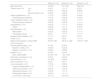
Description of the study groupsDuring the inclusion period nine patients started receiving bevacizumab treatment. Those patients completed at least three treatment cycles (induction phase -36 weeks-) (group A). Mean patient age at the start of treatment was 70 ± 8.4 years; four (44.4%) were female and four (44.4%) presented a ENG gene mutation. All the patients presented epistaxis and had some degree of gastrointestinal involvement, while seven patients (77.8%) had liver involvement of varying degrees and different types. Despite this, the indication for treatment with bevacizumab was for refractory clinical symptoms of HOHF due to liver involvement (without ischaemic cholangitis and not indicated for liver transplant) in two patients (22.2%) while the rest were indicated for severe anaemia due to gastrointestinal involvement. Both patients with HOHF presented liver involvement with predominantly arteriovenous fistulas, hypertrophy of the hepatic artery, and CI of 4.5 and 5.3 (L/min/m2). In the same cohort, we obtained nine control subjects with HHT not indicated for treatment with bevacizumab and matched as previously described (group B). Similarly, we selected nine control subjects without HHT (group C), matched for age and sex. Table 1 describes the baseline clinical characteristics and treatments for the three patient groups. We detected statistically significant differences in haemoglobin concentrations between group A and groups B and C, but not between the latter two. We also found statistically significant differences in the ESS between groups A and B.
Clinical characteristics of the three study groups.
| Group A (n = 9) | Group B (n = 9) | Group C (n = 9) | ||
|---|---|---|---|---|
| Age, mean (SD) | 70 (8.4) | 72.1 (8.6) | 69.8 (8.2) | |
| Tobacco use n (%) | Yes | 0 (0.0) | 1 (11.1) | 0 (0.0) |
| No | 6 (66.7) | 5 (55.6) | 8 (88.9) | |
| Previous tobacco use | 3 (33.3) | 3 (33.3) | 1 (11.1) | |
| Arterial hypertension, n (%) | 7 (77.8) | 5 (55.6) | 4 (44.4) | |
| Pharmacological treatment | 7 (77.8) | 5 (55.6) | 4 (44.4) | |
| Type 2 diabetes mellitus, n (%) | 3 (33.3) | 4 (44.4) | 1 (11.1) | |
| Pharmacological treatment | 3 (33.3) | 4 (44.4) | 1 (11.1) | |
| Dyslipidaemia, n (%) | 2 (22.2) | 1 (11.1) | 5 (55.6) | |
| Statins | 2 (22.2) | 1 (11.1) | 4 (44.4) | |
| Atrial fibrillation, n (%) | 2 (22.2) | 2 (22.2) | 1 (11.1) | |
| Beta blocker | 2 (22.2) | 2 (22.2) | 1 (11.1) | |
| Anticoagulant drugs | 0 (0.0) | 1 (11.1) | 1 (11.1) | |
| Ischaemic heart disease, n (%) | 1 (11.1) | 0 (0.0) | 0 (0.0) | |
| COPD, n (%) | 0 (0.0) | 0 (0.0) | 1 (11.1) | |
| Baseline haemoglobin, median [IQR] | 87 [79−103]* | 135 [115−152]* | 145 [141−164]* | |
| HHT involvement | ||||
| Positive genetic testing, n (%) | 9 (100) | 9 (100) | ||
| Mutation in ENG, n (%) | 4 (44.4) | 4 (44.4) | ||
| Mutation in ACVRL1, n (%) | 5 (55.6) | 5 (55.6) | ||
| Epistaxis, n (%) | 9 (100) | 9 (100) | ||
| ESS, median [IQR] | 5.7 [4.6−7.6]* | 1.5 [1−2.9]* | ||
| Pulmonary involvement, n (%) | 3 (33.3) | 2 (22.2) | ||
| GI involvement, n (%) | 9 (100.0) | 4 (44.4) | ||
| CNS involvement, n (%) | 0 (0.0) | 0 (0.0) | ||
| Liver involvement, n (%) | 7 (77.8) | 5 (55.6) | ||
| Hepatic telangiectasias, n (%) | 7 (77.7) | 4 (44.4) | ||
| Arteriovenous fistulas, n (%) | 3 (33.3) | 3 (33.3) | ||
| Arterioportal fistulas, n (%) | 3 (33.3) | 1 (11.1) | ||
| Portosystemic fistula, n (%) | 2 (22.2) | 1 (11.1) | ||
| CI (L/min/m2), mean (SD) | 3.3 (1.1) | 3.2 (0.74) | ||
Abbreviations: ACVRL1: activin A receptor type II-like 1; HA, hepatic artery; EES: epistaxis severity score; ENG: endoglin; COPD: chronic obstructive pulmonary disease; GI: gastrointestinal; HHT: hereditary hemorrhagic telangiectasia; CI: cardiac index; IQR: interquartile range; SD: standard deviation; CNS: central nervous system.
This is the first study seeking to define the role of various angiogenic and inflammatory biomarkers during bevacizumab treatment in patients with HHT. Their detection will provide objective and reproducible data that may help guide anti-angiogenic treatment with bevacizumab, with a new, especially useful approach during the maintenance phase. We selected biomarkers with more evidence or specific relevance in HHT and other vascular diseases, which are also easily analysed in plasma or serum.23 Similarly, the study allowed us to compare the baseline situation of these biomarkers in patients with HHT with and without indication for bevacizumab, providing prognostic information regarding possible differences between groups. Lastly, we were able to compare the concentrations of these biomarkers in patients with and without HHT, with a potential role in aiding diagnosis in patients with suspected HHT. This was done with an eye to consolidating personalised or precision medicine to maximise the likelihood of individual therapeutic efficacy and reduce to the extent possible any toxicity risk of the treatment used.21–23 The obtained results will also enable researchers to formulate hypotheses for application in other settings, such as blood loss anaemia. Having clinical and laboratory data for three groups also allowed us to analyse the correlation of these biomarkers with a specific underlying disease or organ involvement due to HHT or based on haemoglobin levels. As such, compared to the other two groups, the lower levels of haemoglobin and higher EES score that HHT patients receiving bevacizumab (group A) present will be particularly useful.
The evaluation of biomarkers between patients with HHT and in relation to their severity has been studied in the context of VEGF and adrenomedullin. As for VEGF, Cirulli et al. initially observed elevated concentrations of this biomarker in patients with HHT compared to healthy controls, although later results did not find these differences.26–28,36,37 In the case of adrenomedullin, higher concentrations in blood and greater tissue expression in skin telangiectasia biopsies were found in patients with HHT compared to healthy controls.24 Lower ANGPT2 and sENG concentrations and higher pentraxin 3 and SDF-1 concentrations were also observed in HHT patients compared to control subjects.27,28,38 No differences were found in the other biomarkers studied, and for most, no such comparison has been described.23
One of the limitations of this study was that the selection of biomarkers was arbitrary. Despite this, the selection was very broad, including a large number of biomarkers. In addition, because it was an exploratory study, the magnitude of the potential differences of biomarkers between the three groups is unknown, making it impossible to calculate a sample size. As with other exploratory studies, this was based more on the viability of the proof of concept than on statistical power. Another issue could be the technique and timing of sampling. However, repeating the measurements in the three cycles of the induction phase and in the maintenance phase of group A patients, and twice in group B patients, ensure a more realistic reflection of the dynamics of these biomarker concentrations and contributes to avoiding potential intrapersonal variations during the course of the disease. On the other hand, since it was a single-centre study, patient selection could have been conditioned by the centre’s own characteristics. Therefore, the results should be interpreted with caution and considered as an opportunity to formulate hypotheses. These results will need to be validated in future multicentre studies specially designed for such a purpose and with a larger sample size.
There is a need to optimise and individualise monitoring for bevacizumab therapy in patients with HHT. Incorporating angiogenesis- and inflammation-related biomarkers measurement into routine clinical practice could help meet this need. The resulting data could help formulate new hypotheses to study the clinical pathophysiological and therapeutic benefit of specific biomarkers.
Consent to the publication of clinical dataThe patients involved in this study signed an informed consent form regarding the sharing of their clinical data, which has been anonymised via an alphanumeric identification code. The informed consent form for patients with hereditary haemorrhagic telangiectasia and the transfer of biological samples and clinical data was approved by the Research Ethics Committee for Biomedical Research Projects at Bellvitge University Hospital (Barcelona, Spain; registry number: 2023/1377).
Ethical considerationsThis study was approved by the Research Ethics Committee for Biomedical Research Projects at Bellvitge University Hospital (Barcelona, Spain; registry number: PR061/23).
FundingThe study was funded via the “Ayudas a la investigación FEMI” project granted by the Spanish Internal Medicine Foundation (FEMI), as well as projects PI20/00592 and PI23/00164 from the Carlos III Health Institute (ISCIII) and co-financed by the European Union.
Availability of data and materialsThe data that support the study findings are available upon reasonable request made to the corresponding author.
The authors declare that they do not have any conflicts of interest.
We would like to extend specific thanks to the patients included in the study and to the collaboration from Biobank HUB-ICO-IDIBELL (PT20/00171) integrated in the Carlos III Health Institute Biobanks and Biomodels platform and the Xarxa de Bancs de Tumors de Catalunya (XBTC). We would also like to thank the Bellvitge University Hospital Research Committee for awarding us with a “Beca de investigación posresidencia del Hospital Universitari de Bellvitge (2022-2024)” (Bellvitge University Hospital Post-Residence Research Grant) as it was essential to this study. With institutional support from the CERCA Programme/Government of Catalonia.