Incidentalomas of the thyroid gland are frequently observed in oncological patients undergoing FDG PET/CT imaging for staging or treatment response assessment. This study aims to investigate the utility of SUVmax and ADC values measured by PET/MRI in distinguishing between benign and malignant thyroid nodules.
Materials and methodsWe selected 108 patients (72 females, 36 males; mean age 54 ± 12 years) who underwent routine oncological FDG PET/CT scans for staging or treatment response assessment, with nodule sizes greater than 1 cm. A one-bed neck PET/MRI scan followed the whole-body PET/CT. SUVmax values were measured, and ADC maps were created using DWI with b factors of 50 and 1000s/mm². SUVmax and ADC values were correlated with FNAC results.
ResultsFNAC results revealed 76 (70.4%) benign and 32 (29.6%) malignant nodules among the 108 patients. The mean SUVmax of malignant nodules was significantly higher than that of benign nodules (10.6 ± 8.3 vs. 5.94 ± 5.2, p < 0.001). Similarly, the mean ADC value was lower in malignant nodules compared to benign ones (1.4 ± 0.6 × 10ˉ³ mm²/s vs. 1.8 ± 0.4 × 10ˉ³ mm²/s; p < 0.001). A significant but weak correlation was found between FNAC results and mean SUVmax (r = 0.335), as well as a significant weak negative correlation with mean ADC values (r = -0.355). Using a cut-off value of 6 for SUVmax and 1.56 × 10ˉ³ mm²/s for ADC, the sensitivity, specificity, and accuracy for SUVmax were 68.7%, 73.6%, and 72.1%, respectively, while for ADC, they were 71.8%, 69.7%, and 70.4%, respectively. The PET/MRI system demonstrated a relative sensitivity, specificity, accuracy, PPV, and NPV of 90.62%, 51.32%, 62.96%, 43.94%, and 92.86%.
ConclusionThis study is one of the first in the literature to explore the use of FDG PET/MRI, a single-stop device, in distinguishing between benign and malignant thyroid nodules with high sensitivity and NPV.
Los incidentalomas de la glándula tiroides son frecuentemente observados en pacientes oncológicos que se someten a imágenes FDG PET/CT para la estadificación o evaluación de la respuesta al tratamiento. El objetivo de este estudio es investigar la utilidad de los valores de SUVmax y ADC medidos por PET/MRI para distinguir entre nódulos tiroideos benignos y malignos.
Materiales y métodosSe seleccionaron 108 pacientes (72 mujeres, 36 hombres; edad media 54 ± 12 años) que se sometieron a estudios rutinarios de FDG PET/CT con fines oncológicos para la estadificación o evaluación de la respuesta al tratamiento, con nódulos de tamaño superior a 1 cm. Tras el PET/CT corporal completo, se realizó un PET/MRI de cuello de una cama. Se midieron los valores de SUVmax y se crearon mapas de ADC utilizando imágenes ponderadas por difusión (DWI) con factores b de 50 y 1000s/mm². Los valores de SUVmax y ADC fueron correlacionados con los resultados de la citología por aspiración con aguja fina (FNAC).
ResultadosLos resultados de FNAC revelaron 76 (70,4%) nódulos benignos y 32 (29,6%) nódulos malignos entre los 108 pacientes. El SUVmax promedio de los nódulos malignos fue significativamente mayor que el de los nódulos benignos (10,6 ± 8,3 frente a 5,94 ± 5,2, p < 0,001). De manera similar, el valor promedio de ADC fue inferior en los nódulos malignos en comparación con los benignos (1,4 ± 0,6 × 10ˉ³ mm²/s frente a 1,8 ± 0,4 × 10ˉ³ mm²/s; p < 0,001). Se encontró una correlación significativa pero débil entre los resultados de FNAC y el SUVmax promedio (r = 0,335), así como una correlación negativa débil pero significativa con los valores promedio de ADC (r = -0,355). Utilizando un valor de corte de 6 para el SUVmax y de 1,56 × 10ˉ³ mm²/s para el ADC, la sensibilidad, especificidad y precisión para el SUVmax fueron 68,7%, 73,6% y 72,1%, respectivamente, mientras que para el ADC fueron 71,8%, 69,7% y 70,4%, respectivamente. El sistema PET/MRI demostró una sensibilidad, especificidad, precisión, valor predictivo positivo (PPV) y valor predictivo negativo (NPV) relativos de 90,62%, 51,32%, 62,96%, 43,94% y 92,86%.
ConclusiónEste estudio es uno de los primeros en la literatura en explorar el uso del FDG PET/MRI, un dispositivo de diagnóstico único, para distinguir entre nódulos tiroideos benignos y malignos con alta sensibilidad y un elevado valor predictivo negativo (NPV).
Article
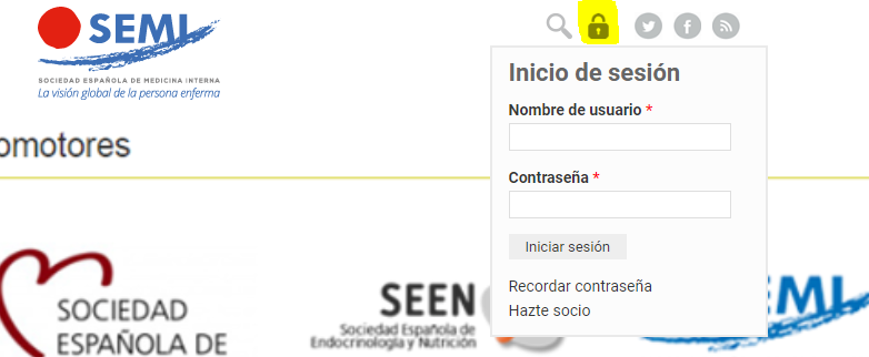
Diríjase desde aquí a la web de la >>>FESEMI<<< e inicie sesión mediante el formulario que se encuentra en la barra superior, pulsando sobre el candado.

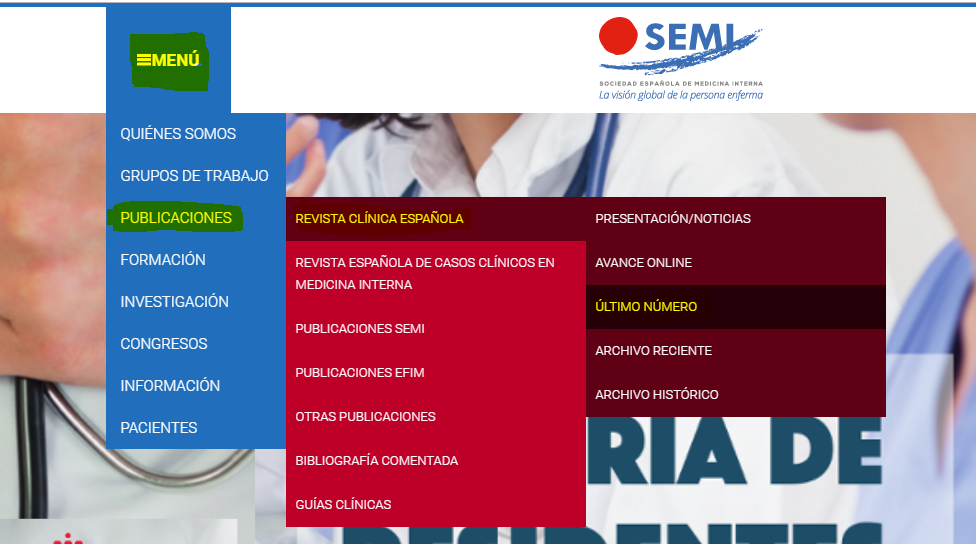
Una vez autentificado, en la misma web de FESEMI, en el menú superior, elija la opción deseada.

>>>FESEMI<<<


